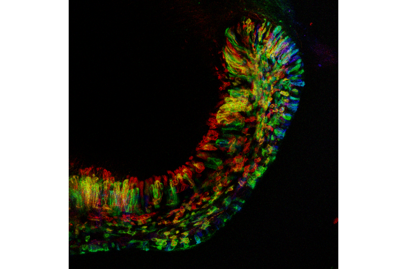
Mouse teeth providing new insights into tissue regeneration

Researchers hope to one day use stem cells to heal burns, patch damaged heart tissue, even grow kidneys and other transplantable organs from scratch. This dream edges closer to reality every year, but one of the enduring puzzles for stem cell researchers is how these remarkable cells know when it's time for them to expand in numbers and transform into mature, adult cells in order to renew injured or aging tissue.
The answer to this crucial decision-making process may lie in a most remarkable organ: the front tooth of the mouse.
Constantly growing incisors are the defining feature of all rodents, which rely on these sharp, chisel-like gnashers for burrowing and self-defense as well as gnawing food. Inside the jaw, a mouse's incisors look more like a walrus's tusks or the teeth of a saber-toothed tiger, with only the sharpened tips showing through the gums at the front of the mouth.
As the front of the tooth gets ground down, a pool of stem cells deep inside the jaw, at the very inner part of the tooth, is constantly building up the back of each incisor and pushing the growing tooth forward—a bit like the lead of a mechanical pencil.
"As we grow older our teeth start to wear out, and in nature, once you don't have your teeth anymore, you die. As a result, mice and many other animals - from elephants to some primates - can grow their teeth continuously," said UC San Francisco's Ophir Klein, MD, PhD, a professor of orofacial sciences in UCSF's School of Dentistry and of pediatrics in the School of Medicine. "Our lab's objective is to learn the rules that let mouse incisors grow continuously to help us one day grow teeth in the lab, but also to help us identify general principles that could enable us to understand the processes of tissue renewal much more broadly."
In a new study, published online April 27, 2017, in Cell Stem Cell, Jimmy Hu, PhD, a postdoctoral researcher in the Klein laboratory, has discovered that signals from the surrounding tissue are responsible for triggering these dental stem cells to leave their normal state of dormancy, hop on the conveyor belt of the growing tooth, and begin the process of transforming into mature tooth tissue.
"We usually think of stem cells responding to chemical signals to start proliferating and differentiating, but here there's an exciting interaction between the physical environment and the cells that can prompt them to meet the demands of the growing tooth," Hu said.
In their study, Hu and colleagues discovered that integrins, proteins that sit in cell membranes and link the internal skeleton of cells to the larger protein scaffolding of the surrounding tissue, trigger a newly described signaling cascade within the stem cells that causes them to begin rapidly multiplying - a process called "proliferation."
It's not clear yet exactly what external signals are responsible for triggering the stem cells to proliferate, the authors say, but they propose that the cells could be detecting that they have moved into a region where the back of the tooth needs to actively produce more cells based on changes in local tissue stiffness or the physical forces pulling and pushing on the cells.
"Our data clearly show that as stem cells move into their designated proliferating space, they ramp up integrin production. These integrins allow the cells to interact with extracellular molecules and become triggered to expand in numbers before eventually producing a large pool of mature dental cells," Hu said.
Of additional interest to the researchers is the fact that both integrins and YAP - one of the molecules involved in the newly discovered integrin-triggered signaling cascade - have previously been implicated in the growth of certain types of tumors, which are thought to share some features of stem cell biology. This finding adds evidence to a growing sense among cancer researchers that interactions between cancer cells and the surrounding tissue may be a key step in triggering tumor growth.
"Integrins and YAP had been implicated in cancer before, but our work connects the two in an organ as opposed to in a Petri dish," Klein said. "Wouldn't it be nice if the same insights that let us learn to grow new tissues in the lab also lead to improved therapies to prevent the growth of tumors in patients?"
More information: Jimmy Kuang-Hsien Hu et al, An FAK-YAP-mTOR Signaling Axis Regulates Stem Cell-Based Tissue Renewal in Mice, Cell Stem Cell (2017). DOI: 10.1016/j.stem.2017.03.023


